A microdiscectomy recovery timeline follows a fairly predictable pattern, but how each phase feels depends on how long the nerve was compressed before surgery and how the body responds afterward. Microdiscectomy is a type of spine surgery and a minimally invasive surgery designed to relieve pressure on the spinal nerves by removing only the herniated portion of the disc, rather than the entire disc as in traditional surgeries. Traditional surgeries often involved removing the entire disc and required larger incisions, which led to more tissue disruption, scarring, and longer recovery times. Microdiscectomy specifically targets the spinal canal to address pain caused by nerve compression from a herniated disc.
Advancements in minimally invasive microdiscectomy techniques have significantly improved recovery experiences, resulting in less postoperative pain and shorter hospital stays. Some patients notice leg pain relief almost immediately. Others improve more gradually, especially when nerve irritation has been present for months.
Understanding what tends to happen week by week makes recovery easier to navigate and helps set realistic expectations.
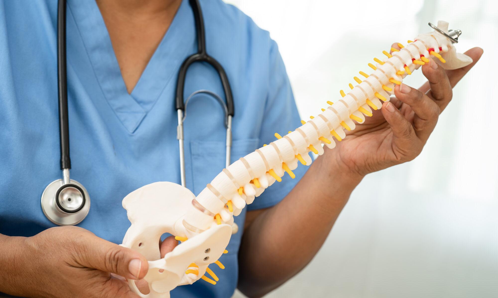
What Is Microdiscectomy and How Does It Work?
Microdiscectomy is a minimally invasive surgical procedure designed to address pain and neurological symptoms caused by a herniated disc pressing on the spinal nerves. During microdiscectomy surgery, the orthopedic surgeon removes the portion of the spinal disc that is causing nerve compression, which often results in significant leg pain, numbness, or weakness. This surgical procedure is typically recommended for patients who have not achieved relief from conservative treatments such as physical therapy, pain medication, or rest. Because microdiscectomy targets only the damaged part of the disc and uses small incisions, it generally allows for a quicker recovery and less disruption to surrounding tissues compared to more invasive spinal surgeries.
Many patients experience rapid pain relief and improved mobility, making microdiscectomy a highly effective option for those suffering from persistent symptoms due to a herniated disc.
What to Expect in the First Few Days
After microdiscectomy surgery, patients are monitored in a recovery room or recovery area for a few hours to ensure they are stable before being discharged. The first few days focus on pain control and gentle movement. Postoperative pain is common, especially around the surgical incision, and patients may experience pain that is different from their preoperative symptoms. Surgical soreness in the lower back is common, even when leg symptoms improve quickly. Many patients describe a different type of discomfort than before surgery, more localized and less sharp.
Walking usually starts early. Short, frequent walks support circulation and reduce stiffness. Patients are advised to avoid prolonged sitting and heavy lifting during the first few days to prevent strain on the healing spine and promote proper recovery. Sitting tends to feel limited at first, and most patients find they need to change positions often.
Nerve-related symptoms do not always disappear immediately. It is normal to experience pain or discomfort following surgery, and monitoring and reporting any unusual pain to the healthcare team is important. Tingling or mild discomfort in the leg can continue as the nerve settles, even when pressure has been relieved.
Weeks 1–2: Early Recovery Phase
During the first two weeks, mobility improves steadily. Walking distance increases, and basic daily activities become more manageable. Sitting tolerance often improves, though extended periods can still trigger discomfort, so prolonged sitting should be minimized to support healing.
Some patients feel significant relief in this phase. Others notice symptoms come and go. That fluctuation is common and usually reflects the nerve adjusting rather than a setback.
Post microdiscectomy restrictions still matter here. Patients should avoid bending, twisting, and strenuous activities, as these actions can lead to further injury or complications during the healing phase. Lifting should also remain limited. The focus stays on controlled movement rather than pushing activity too quickly.
Following postoperative instructions, including activity restrictions, wound care, and medication management, is crucial to ensure a smooth and optimal recovery.
Weeks 3–6: Gradual Return to Activity and Physical Therapy
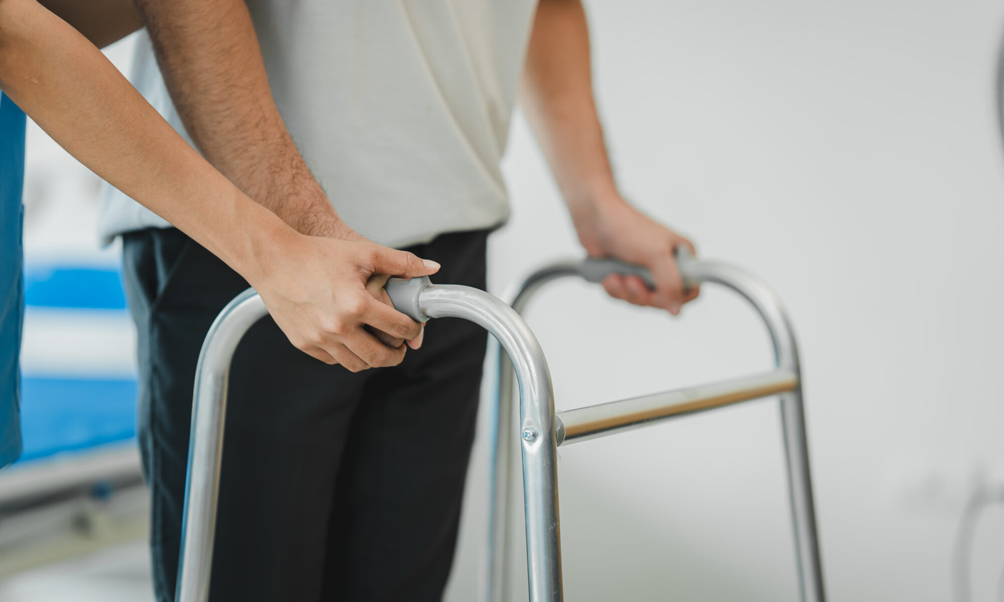
By this stage, many patients begin to feel more stable in their movement. Walking becomes easier, and daily routines start to feel closer to normal. Sitting and standing for longer periods becomes more tolerable. Gentle stretching and low-impact exercises are important at this point to support the healing of soft tissues and promote flexibility.
Physical therapy typically starts about 2 to 4 weeks after surgery, once the incision has healed and pain has decreased. Attending physical therapy is a key part of recovery, as physical therapists guide patients through exercises to gradually increase activity levels, strengthen core and back muscles, and improve posture. Physical therapy also helps patients relearn safe movement patterns, such as proper lifting, bending, and twisting, to avoid re-injury and support long-term spinal health.
Return to work depends on the type of job. Those with sedentary roles may return sooner, often with modifications. Jobs that involve physical activity typically require more time.
It is common to still notice mild tightness or occasional nerve sensations. These do not always indicate a problem. Nerve tissue heals slowly, and improvement can continue over several weeks.
Weeks 6–12: Strength and Stability Phase
The focus shifts toward rebuilding strength and improving spinal stability. Core stability becomes a key focus of physical therapy during this phase, and attending physical therapy sessions supports long-term recovery and spinal health. At this point, many patients resume more regular activity, though high-impact or heavy lifting may still be limited. Most patients can resume light activities within 2 to 4 weeks after surgery, progress to moderate activities by 6 to 8 weeks, and return to their usual daily routines by 8 to 12 weeks post-operation.
Many patients can start driving within 1-2 weeks after surgery, depending on their recovery and pain levels, and are typically allowed to return to low-impact sports by 6 months if they are pain-free.
Lingering symptoms often continue to improve during this period, and patients may experience sensations such as “pins and needles” as nerves regenerate post-surgery. By the three-month mark, many patients can return to near-normal activities, including high-impact sports and heavy lifting, provided they experience no lingering pain or stiffness. Recovery from a lumbar microdiscectomy generally spans several months, with full recovery often occurring between 3 to 6 months post-surgery, although some patients may continue to improve for up to a year. Complete recovery of nerve function and core strength could take up to 3 to 6 months, and nerve healing can be delayed by chronic health conditions like diabetes or by factors such as smoking.
How long it takes to recover from microdiscectomy varies. The general timeline provides a guide, but individual progress depends on factors such as pre-surgical condition, activity level, and overall health.
Managing Pain and Discomfort
Following microdiscectomy surgery, it’s normal to experience some pain and discomfort as the body begins to heal. Most patients find that pain can be managed effectively with pain medication, but it’s important to use these medications exactly as prescribed by your healthcare provider. While narcotic pain medication may be used in the first few days, many patients transition to non-narcotic pain medication or over-the-counter options as their discomfort lessens. In addition to medication, gentle exercises such as short walks can help reduce stiffness and promote circulation. Applying ice or heat to the lower back, taking regular breaks to rest, and attending physical therapy sessions can also support pain management and speed up recovery. Physical therapy is especially valuable for teaching safe movement patterns and helping patients regain strength and flexibility after surgery.
Incision Care and Wound Healing
Proper care of the incision site is essential for smooth healing after microdiscectomy surgery. Patients should keep the incision site clean and dry, following their healthcare provider’s instructions for bathing and changing dressings. It’s important to avoid submerging the incision in water, such as in a bathtub or swimming pool, until it is fully healed. Applying topical antibiotics as directed and monitoring the area for any changes can help prevent infection. If you notice increased redness, swelling, or pain at the incision site, contact your healthcare provider promptly. By following these guidelines, patients can support the healing process and reduce the risk of complications.
Signs of Infection or Complications
Although microdiscectomy surgery is considered safe, it’s important to be aware of potential signs of infection or complications. Watch for symptoms such as increased pain, redness, or swelling at the incision site, as well as fever, chills, or nausea. Additionally, new or worsening numbness, tingling, or weakness in the legs may indicate nerve involvement. If any of these symptoms occur, notify your healthcare provider right away. Early recognition and treatment of complications are key to ensuring a successful recovery and preventing more serious issues. Staying alert to changes in your condition helps you take an active role in your recovery process.
When Recovery Feels Slower Than Expected
Not every recovery follows a smooth upward trend. Some patients notice periods where progress seems to stall or symptoms briefly return.
This often relates to nerve healing rather than a new issue. A nerve that has been irritated for a long time does not settle immediately once pressure is removed. Sensations like tingling, sensitivity, or mild discomfort can persist while healing continues. In some cases, however, symptoms may recur if the same disc herniates again, which may require further evaluation or treatment.
Gradual improvement over time matters more than day-to-day changes. Patterns that worsen steadily or fail to improve should be evaluated, but short-term fluctuations are common.
When to Follow Up With a Spine Specialist for a Herniated Disc
Certain symptoms should prompt follow-up:
- worsening leg pain after initial improvement
- persistent numbness or weakness
- difficulty with basic movement that does not improve
Evaluation at that point focuses on how recovery is progressing and whether further intervention is needed.
At ISSI, follow-up care centers on how symptoms present in real movement, not just imaging findings. That approach helps guide next steps and keeps recovery aligned with function, not just timelines. Adhering to postoperative guidelines, including activity restrictions and attending all follow-up appointments, is essential to support recovery and prevent complications.
Promoting Long-Term Spinal Health
Maintaining spinal health after microdiscectomy surgery is crucial for preventing future problems and supporting a full recovery. Patients are encouraged to maintain a healthy weight, engage in regular low-impact exercises, and practice proper posture throughout daily activities. Attending physical therapy sessions can help strengthen core muscles, improve flexibility, and teach strategies to avoid re-injury. It’s important to avoid heavy lifting, bending, or twisting, especially in the early stages of recovery, as these movements can strain the spine. Working closely with your healthcare team allows you to develop a personalized plan for long-term spinal health, ensuring you can return to normal activities safely and reduce the risk of future back issues. By making these habits part of your routine, you can protect your spine and enjoy lasting benefits from your microdiscectomy surgery.
A Realistic View of Recovery
A microdiscectomy recovery timeline gives patients a general sense of what to expect after surgery, but recovery does not look exactly the same for everyone. Some people notice rapid relief from leg pain within days, while others continue improving gradually over several months as irritated nerves heal and strength returns. Paying attention to movement restrictions, attending physical therapy, and allowing the body enough time to recover all play an important role in long-term results.
Most patients are able to return to normal activity over time, especially when recovery is approached with consistency and realistic expectations. The goal is not simply pain relief, but restoring function, stability, and confidence in movement without placing unnecessary stress on the spine again.
If you are considering surgery for a herniated disc or have questions about your recovery after microdiscectomy surgery, the team at International Spine and Sports Institute (ISSI) can evaluate your condition and discuss treatment and recovery expectations based on your symptoms, imaging, and activity level.